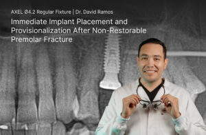
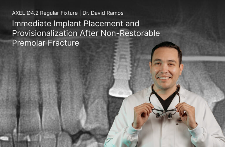
A previously endodontically treated maxillary right second premolar (#4) presented with a structural fracture and was deemed non-restorable. Dr. David Ramos performed an atraumatic extraction followed by Dentis Guide-guided immediate placement of an AXEL 4.2 × 12 mm implant in a prosthetically driven position.
Simultaneous bone grafting and immediate provisionalization were performed to support preservation of the extraction site and surrounding tissue architecture, with the provisional restoration kept out of occlusion during the initial healing phase.
Case Background
A 44-year-old female patient presented with a previously endodontically treated maxillary right second premolar (#4) with a structural fracture, rendering the tooth non-restorable.
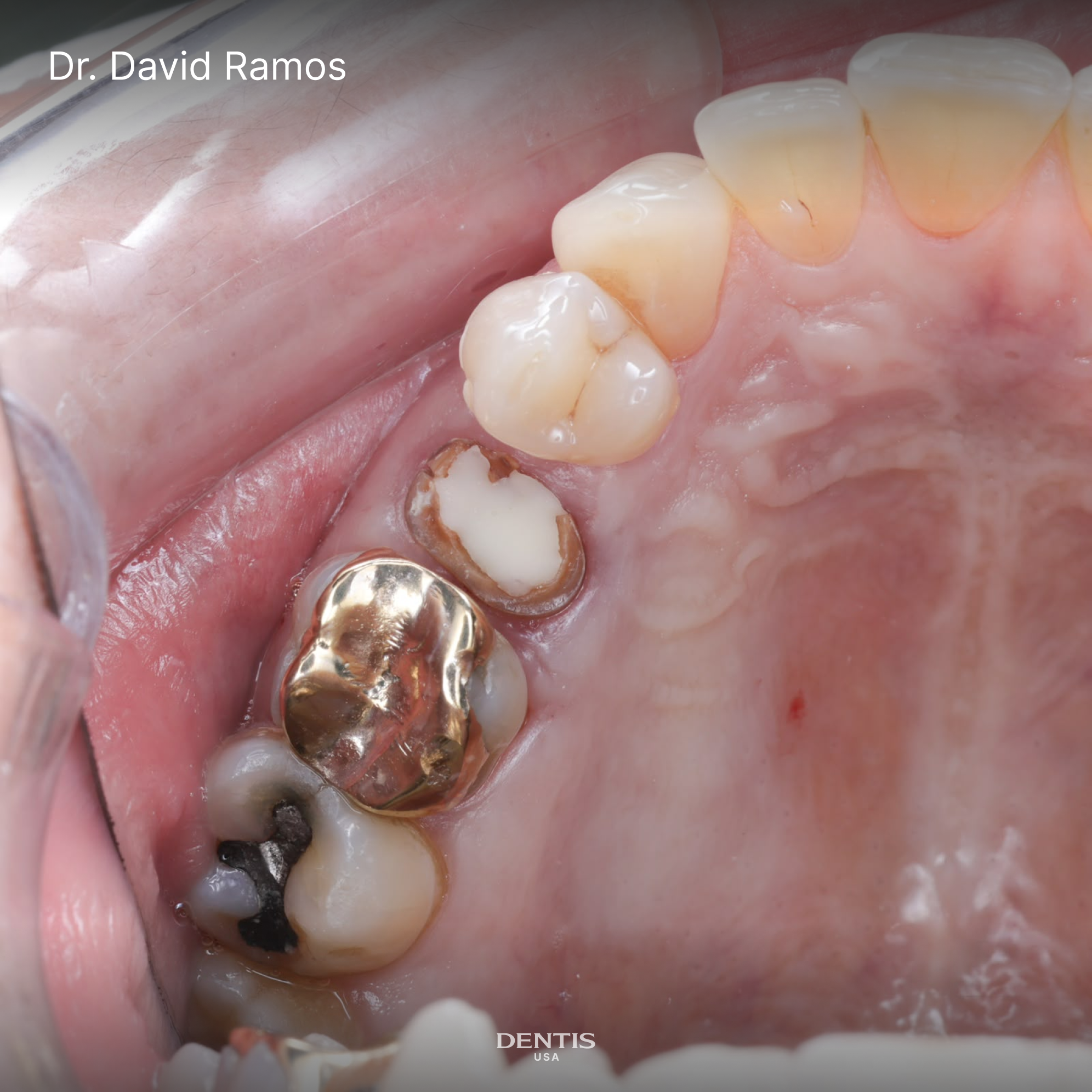
Image displays structural non-restorable fracture.
Following evaluation, Dr. Ramos elected to proceed with immediate implant placement using the Dentis Guide, along with simultaneous bone grafting to maintain the extraction site and support favorable tissue healing and restoration.
Initial Assessment & Treatment Plan
Clinical evaluation revealed D3 bone density. Following assessment, immediate guided implant placement was selected to preserve the extraction site, support the surrounding tissue architecture, and reduce overall treatment time.
Surgical Procedure
1. Tooth Extraction
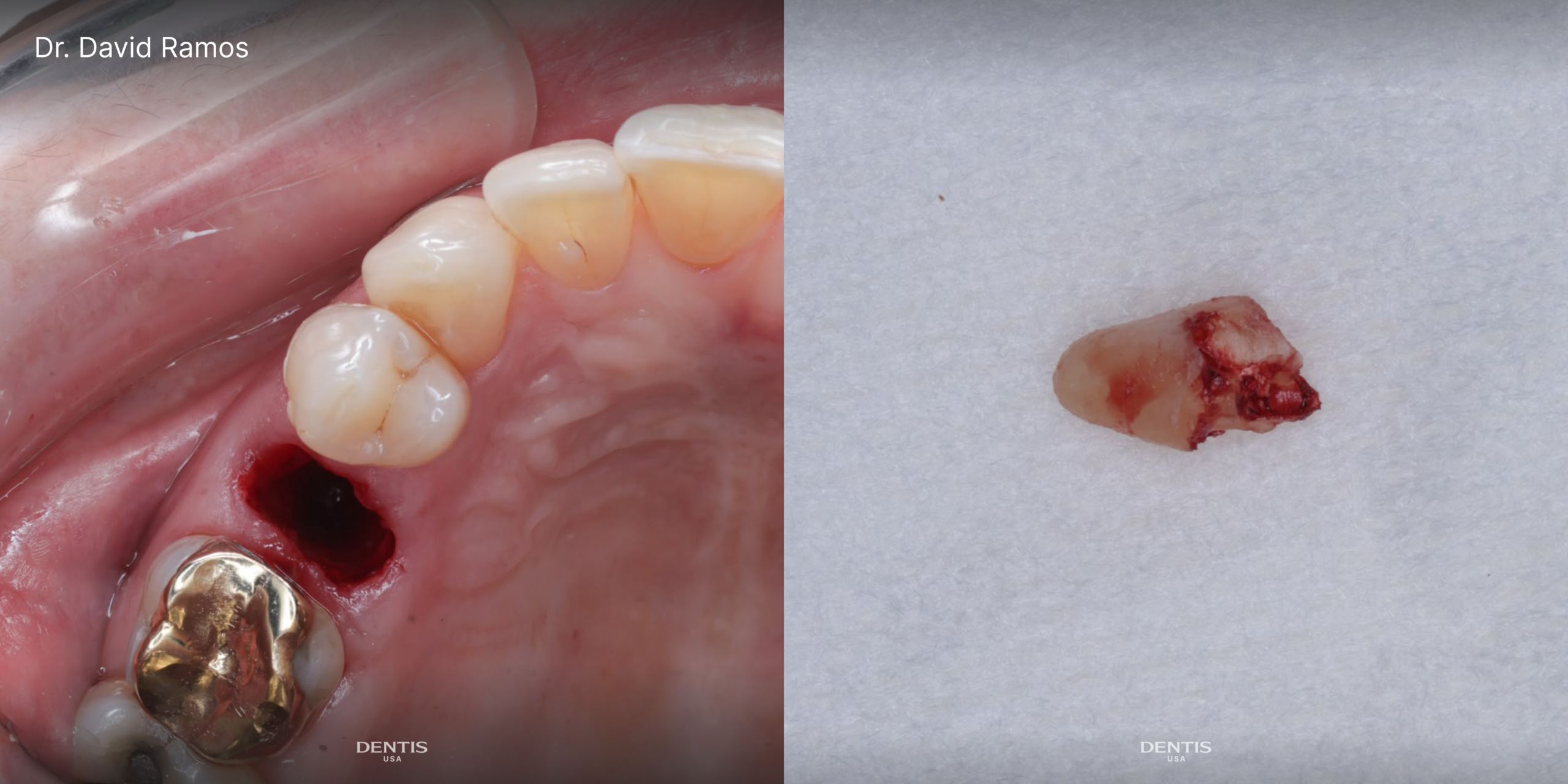
Images show extraction site and extracted tooth.
The tooth was atraumatically extracted, followed by guided immediate placement of an AXEL 4.2 × 12 mm implant using the Dentis Guide. The implant was positioned according to the prosthetically driven treatment plan to support the desired restorative outcome.
2. Guided Implant Placement
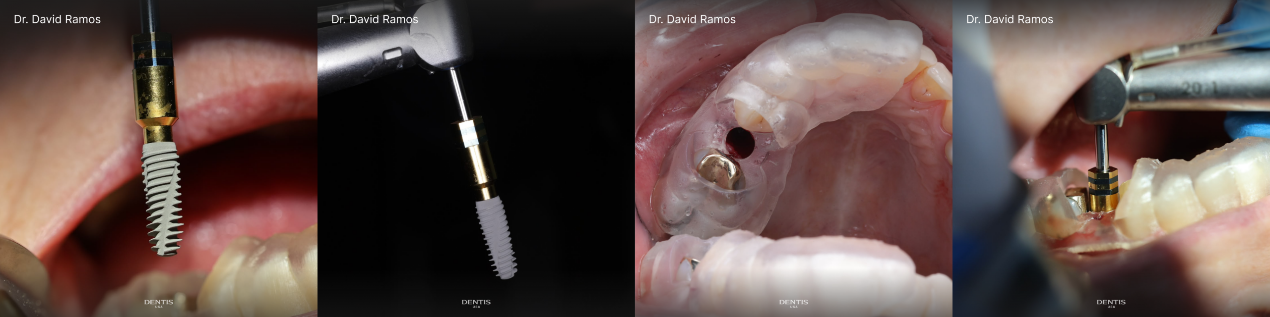
AXEL Implant 4.2 x 12mm placement with Dentis Guide.
A guided immediate placement of an AXEL 4.2 × 12 mm implant was performed using the Dentis Guide System. The implant was positioned according to the prosthetically driven treatment plan to support the desired restorative outcome.
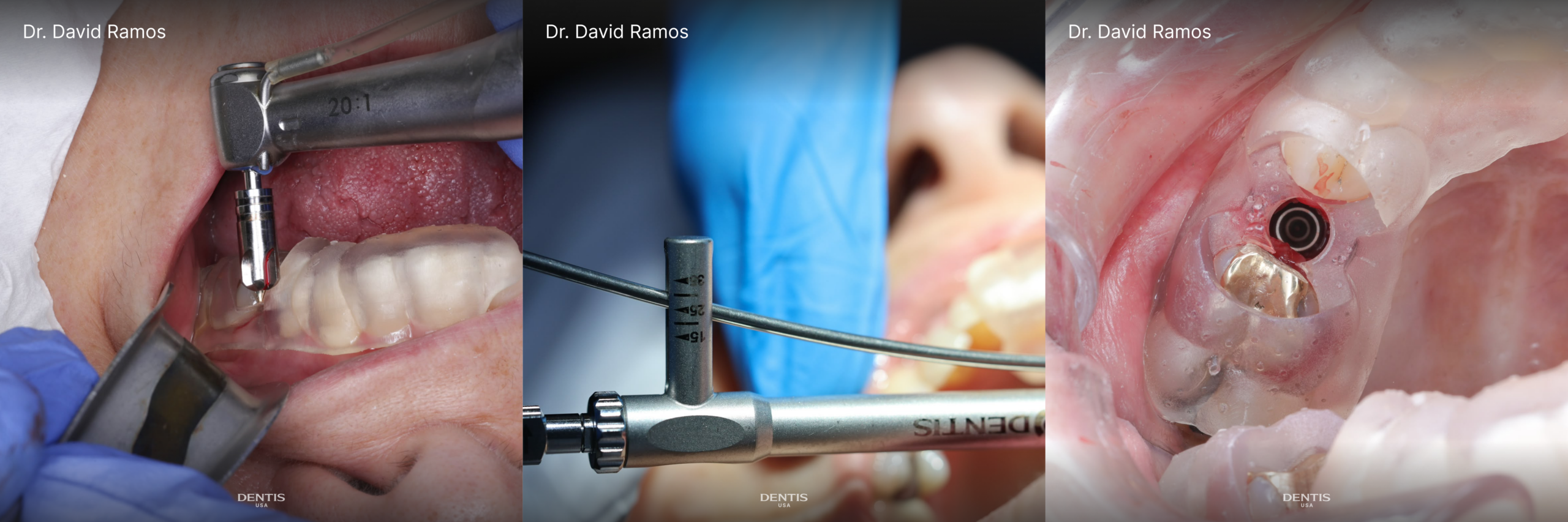
3. Bone Grafting
Bone grafting was performed with allograft simultaneously to preserve the surrounding hard-tissue architecture and support healing of the extraction socket.
4. Provisional Restoration
Following implant placement, an intraoral scan was performed to capture the implant position and surrounding anatomy for fabrication of the provisional restoration.
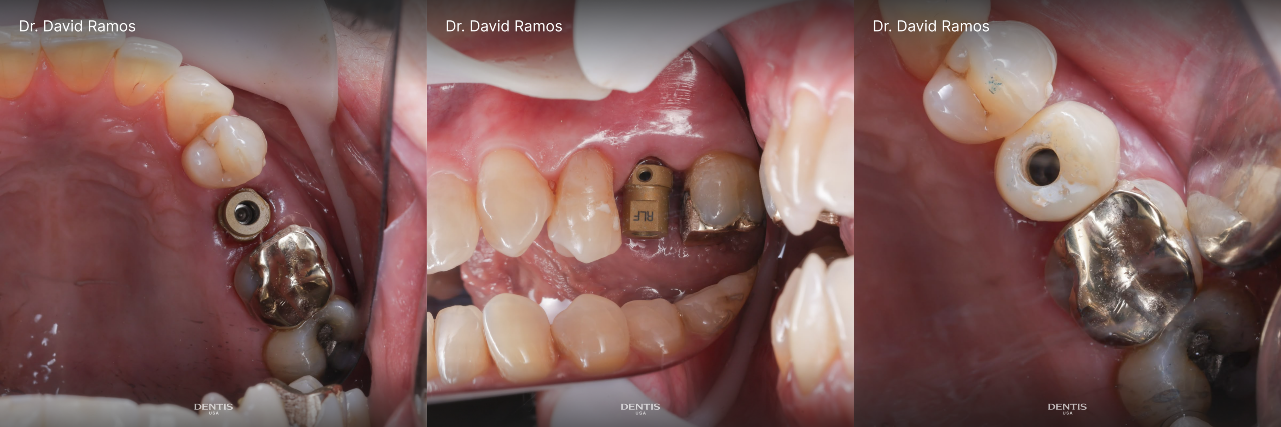
Images show ScanBody inserted after AXEL Implant placement.
The provisional restoration was adjusted completely out of occlusion to protect the implant during the initial healing phase while maintaining the intended emergence profile and surrounding soft-tissue architecture.
Final Result & Follow-Up
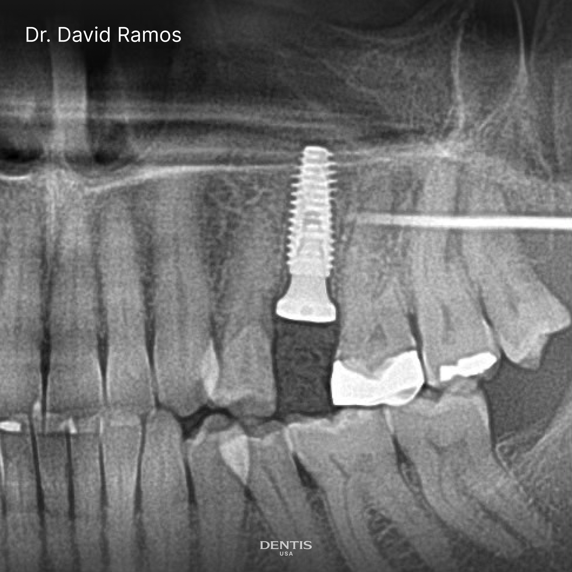
Final x-ray image post surgery.
For more information on this implant, AXEL, feel free to check out our dentishop or contact us to connect with a local sales representative.